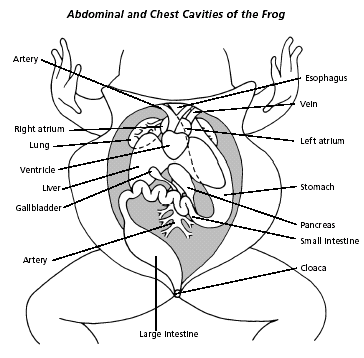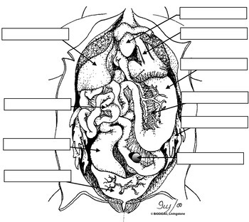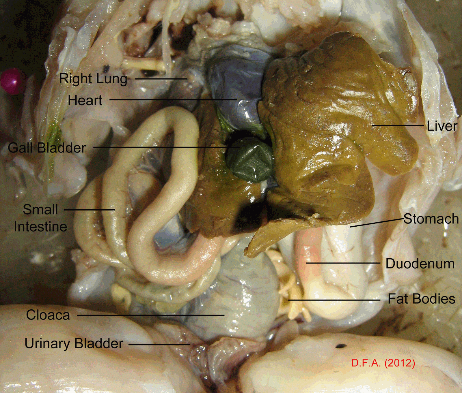
To learn and practice dissection technique Materials and Equipment Dissection tray Dissection kit Preserved. To assess the function of structures from observing the actual anatomy of the organism 5. To locate the structures, organs and systems of the frog 4. I also show them a photo album of frogs we have looked at over the years and help them to compare the photos of the frogs to diagrams of their anatomy. Frog Dissection Diagram Labeled Worksheet - Wiring Diagram Source . To become acquainted with the internal anatomy of the frog 3. We trace the path of food from the esophagus to the stomach, duodenum, ileum, and finally the large intestine (cloaca). socket Gullet Glottis Tongue Labeled Diagram for Lab 19 Frog Dissection doc Digital Frog 2 workbook sample May 2nd, 2018 - diagram and label the parts of the heart Arteries The. Mouthparts of the Frog Grafton Public Schools. Frog Sandwich Labels Pleasanton Unified School District. Beginning anatomy students often make the mistake of trying to memorize drawings or photos rather than attempt to make a mental diagram of how the parts fit together. Cat Dissection Diagrams To Label ImageResizerTool Com. CLICK ON THE DESCRIPTIONS BELOW TO VIEW PICTURES OF THE FROG DISSECTION. I usually do this practice the day after students have dissected the frog to help them conceptualize how the structures fit together. Opening in the mouth that leads to the tympanic membrane on the outside of the frog's head. ( The worksheet could be modified to not include it should students need a greater challenge. Terms in this set (16) Two bony plates on the roof of the frog's mouth that help hold prey before swallowing. The main structures of the abdominal cavity are shown on this image and students practice identifying them using the included word bank.

The appendix in humans is the evolutionary remains of a larger cecum in human ancestors.This worksheet is a supplement to the frog dissection activity where students examine a preserved specimen. The cecum is large in herbivores but much of it has been lost during evolution in humans. Small Intestine - The principal organ of. It houses bacteria used to digest plant materials such as cellulose. Functions of the Internal Anatomy of a Frog: Stomach - Stores food and mixes it with enzymes to begin digestion. The cecum is a blind pouch where the small intestine joins the large intestine. The spleen is an elongate, flattened, brownish organ that extends along the posterior part of the stomach ventral to (above) the pancreas.

Lift the stomach and identify this light-colored organ.

It extends along the length of the stomach from the left side of the body (your right) to the point where the stomach joins the small intestine. The pancreas is located dorsal and posterior to the stomach. Find the bile duct that leads to the small intestine. This structure stores bile produced by the liver. Lift the right lobe and find the gallbladder. Locate the cecum, a blind pouch where the small intestine joins the large intestine. Find the posterior part of the large intestine called the rectum and observe that it leads to the anus. Identify the small intestine and large intestine. Using a probe, trace follow the esophagus to the stomach. You have already seen how the esophagus leads from the pharynx through the neck region. The word “urogenital” refers to an opening that serves both the urinary (excretory) and the reproductive systems.įigure 20. Esophagus, larynx, trachea, bronchus, and lung. Use your pig and also a pig of the opposite sex to identify the structures in the photographs below. Obtain a fetal pig and identify the structures listed in figure 1. Use figures 1–4 below to identify its sex. The pig in figure 1 below has its ventral side up. The pig in figure 1 is lying on its dorsal side. If a structure is anterior to another, then it is closer to the head. The following words will be used to help identify the location of structures. To gain a first-hand knowledge in vertebrate anatomy students are asked to dissect toad or frog at the very beginning. Links to high-resolution, unlabeled photographs are also provided for many of the photographs. As a result, a structure shown in one photograph may look different than the same structure shown in another photograph.Ĭlick on any of the photographs to view enlargements. Several different pig dissections were used to obtain the photographs below. Microsoft Word - Labeled Diagram for Lab 19 - Frog Dissectiondoc Author. The incision can be seen in the first photograph below. Septemin diagram, dissection, frog, worksheet Locate and observe it Note The pancreas can be challenging to find. An incision was made on the side of the neck to enable the injections. Circulatory System Of Frog 2.1 Heart of frog 2.2 Venous system of frog 2.3 Arterial system of frog 3. The arteries have been filled with red latex and the veins with blue. FROG DISSECTION SHOWING :- 1.External Features Of Frog 2.

The fetal pig that you will dissect has been injected with a colored latex (rubber) compound.


 0 kommentar(er)
